Optical Coherence Tomography (OCT) is a new imaging technique based on low coherence measurement. It can perform non-contact and non-invasive tomography of living tissue microscope structures. OCT is an optical analog of ultrasound with higher longitudinal resolution and does not adversely affect organisms like X-rays and RF electromagnetic fields.Therefore, OCT is particularly suitable for samples with high scattering and non-transparent properties, and organisms are such samples.
At present, more and more OCT is applied to the diagnosis of biological tissues, especially the diagnosis of ophthalmology and subcutaneous tissue. Its penetration depth is almost independent of the transparent refractive medium of the eye. It can observe the anterior segment of the eye and display the morphological structure of the posterior segment of the eye. It has a good application prospect in intraocular diseases, especially in the diagnosis, follow-up observation and the treatment effect evaluation of retinal diseases.
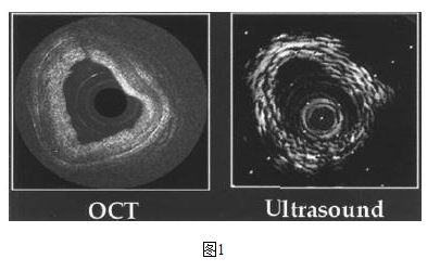
Figure 1 compares the OCT image with the results of the ultrasonic test. Obviously, the resolution of the OCT is higher and the image is clearer.

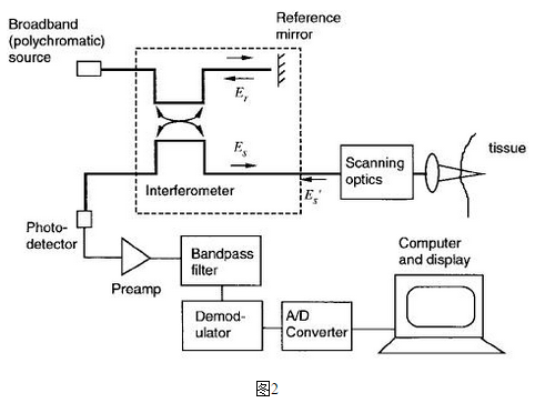
The basic principle of OCT is shown in Figure 2. The basic function is 2Х2 WDM. Both the detection light and the reference light are input into the fiber, and are divided into two parts in the fiber coupler, one part enters the reference arm and one part enters the sampling arm. When the light reflected from the reference arm interferes with the light reflected from the sampling arm, the strongest signal will be obtained on the interference arm detector. Then, by sampling different space points, information about different spaces can be obtained. After filtering and digital-to-analog conversion, the information is converted into a video displayable image. Thus, light is the signal carrier, the optical signal and the resulting information are associated, and the choice of source has a significant impact on the performance of the OCT.

For the choice of OCT light source, there are two points worth noting:
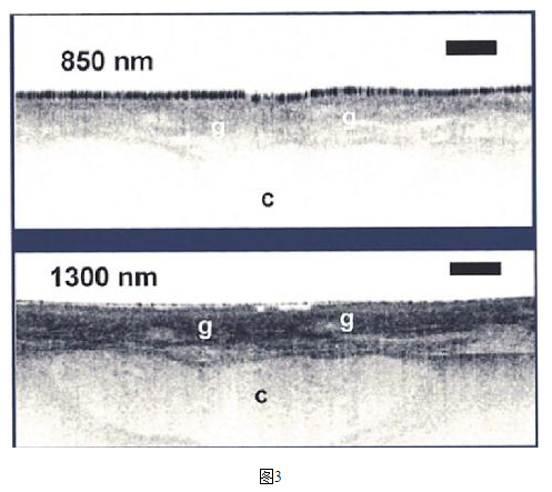
First, human cells have strong scattering of light below 850 nm, and the water in the cells has a high absorption rate for light above 1500 nm. Both of these points are extremely unfavorable for the application of OCT. Therefore, usually the source of the OCT requires a wavelength between 850 and 1600 nm. Figure 3 shows OCT images of laryngeal cartilage at 850 nm and 1300 nm respectively. The image at 1300 nm is clearer.

Second, the longitudinal scanning resolution of the OCT is determined by the coherence length of the source.

Therefore, in order to achieve higher resolution, it is necessary to use a wide-spectrum light source, such as the commonly used super luminescent diode (SLD), and the super-continuous spectrum of the popular femtosecond laser pump.
SLD, the spectral width is typically 40 nm and can reach up to 100 nm. With the development of ultrafast laser technology and the emergence of photonic crystal fibers, it is now possible to use the super-continuous spectrum of the femtosecond laser pump with a spectral width of several hundred nanometers. The emergence of photonic crystal fibers will bring a revolutionary change in the field of optical information technology. In the super-continuum generation process of photonic crystal fibers, femtosecond lasers have more intense dispersion effects and nonlinear effects than picosecond and nanosecond lasers, so that a larger spectral width can be obtained. For example, Germany's Menlosystems uses its femtosecond fiber laser as a pump source to produce a super-continuum of about 400 nm.
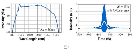
We take Menlosystems' Yb-doped fiber laser (center wavelength 1030nm) as an example, in the mode-locked state, it has a broad spectrum of 56 nm (FWHM) and a pulse width of 58 fs. This allows it to achieve greater dispersion and nonlinear effects, resulting in greater spectral broadening through nonlinear fibers.
